Figure 3.
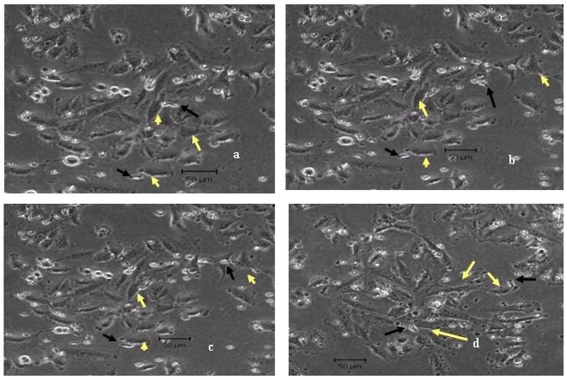
Time-lapse imaging of untreated MCF-7 cells co-cultured with Vγ9Vδ2 T cells. Untreated MCF-7 cells were co-cultured with Vγ9Vδ2 T cells at a 1:2 ratio for 4 hours at 37˚C. Snapshots of continuous time-lapse imaging on a LSM510 Meta Zeiss confocal microscope taken during the last 1 hour are shown. The images were taken at 30 seconds interval (a-d). Vγ9Vδ2 T cells (black arrows) were unable to lyse the tumor cells (yellow arrows) even at the end of 4 hours (d). Representative images from one of four independent experiments are shown.

