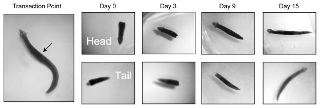
Figure 1. Transection of Planaria.
The image in the left panel shows an intact planarian with head facing up. The arrow indicates the transection point. The adjacent panels show representative images of heads and tails from transected Planaria at days 0, 3, 9 and 15. The completely regenerated Planaria at day 15 offers visible details of the resultant head and tail. The images have been enlarged 16X to show detail.

