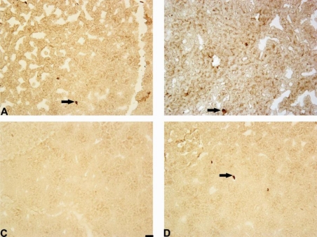
Figure 8.
Photomicrograph of Notch-3-positive cells in the striatum. Immunohistochemical staining, magnification × 200. Internal scale bars = 50 μm. (A) Saline; (B) mildronate at 50 mg/kg; (C) 6-OHDA; (D) M50 + 6-OHDA. The administration of 6-OHDA decreased the number of Notch-3-positive cells in the rat striatum, but the co-administration of mildronate at 50 mg/kg and 6-OHDA increased the number of Notch-3-positive cells to values similar to those observed in controls. Arrows indicate positively stained cells.

