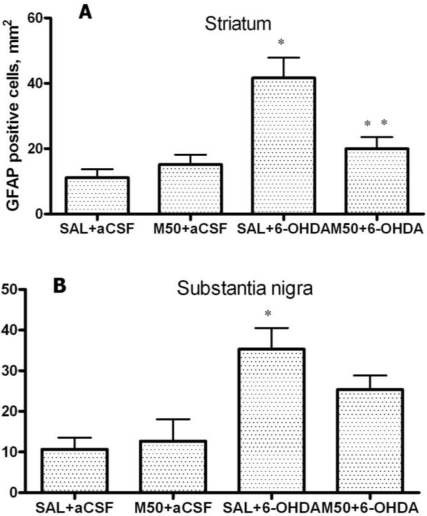
Figure 9.
(A) The number of GFAP-positive astrocytes in the 6-OHDA-lesioned striatum; and (B) substantia nigra. Immunohistochemical examination of rat tissue using a GFAP antibody. Saline (SAL, 1 mL/kg) and mildronate at 50 mg/kg (M50) and 100 mg/kg (M100) were administered intraperitoneally two weeks prior to the injection of 6-OHDA or artificial cerebrospinal fluid (aCSF); 6-OHDA injection in mildronate-treated rats: M50 + 6-OHDA and M100 + 6-OHDA. Striatum: * p = 0.001, SAL + 6-OHDA vs. SAL + aCSF; ** p = 0.01, M50 + 6-OHDA vs. SAL + 6-OHDA; S. nigra: * p = 0.002, SAL + 6-OHDA vs. SAL + aCSF; unpaired t-test. Number of animals per group (n = 8).

