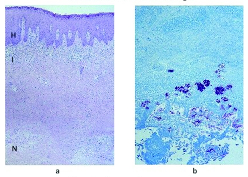
Figure 1.
a, Hematoxylin and eosin stain of a lesion specimen showing definitive Buruli ulcer disease in the preulcerative stage (original magnification 50x). Notice the psoriasiform epidermal hyperplasia (H), superficial dermal lichenoid inflammatory infiltrate (I), and necrosis of subcutaneous tissues (N). b, Ziehl-Neelsen stain of the same nodule, showing abundant colonies of acid-fast bacilli in the necrotic subcutaneous tissues (original magnification 100x).

