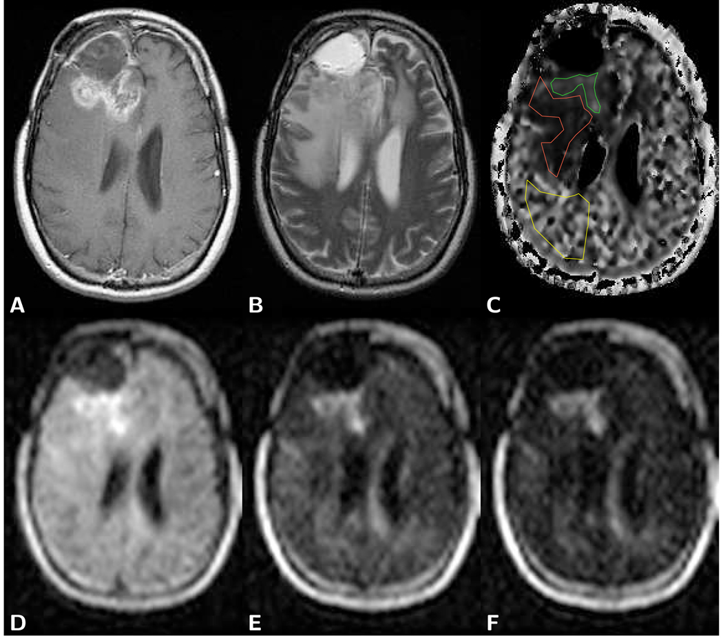
Figure 2.
Axial brain images of a frontal glioblastoma with postoperative cyst formation in a 38 year old male patient. A) T1-weighted post-contrast spin-echo image (TR 600 ms/TE 25 ms) predominantly shows the tumor marginal area and not the solid part of the tumor. B) T2-weighted spin-echo image (TR 3,600 ms/TE 98 ms) shows a bright cyst and edema-related hyperintensity. C) Computed slow diffusion component size image with overlaid ROIs for tumor (green, fs=0.23), edema (red, fs=0.11), and normal appearing brain tissue (yellow, fs=0.35). D) LSDI image with a b-factor of 1,000 s/mm2 shows hyperintensity within the tumor. E) LSDI image with a high b-factor of 3,000 s/mm2 also shows hyperintensity within the tumor. F) Very high diffusion-weighted LSDI image with a b-factor of 5,000 s/mm2 exhibits signal above the noise threshold for all tissues. Extraordinary high residual signal, despite high diffusion weighting, is observed in the solid part of the tumor. LSDI images were all obtained with a single diffusion encoding direction and the following scan parameters: 64×48 imaging matrix interpolated to a 25×192 matrix, 220 mm×165 mm field of view, 7.3 mm slice thickness, 2,040 ms TR, 94 ms TE, 16 b-factors. The diffusion values measured with conventional diffusion imaging (b=1,000 s/mm2) were 1.10 µm2/ms for tumor and 0.74 µm2/ms for normal appearing white matter.

