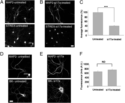
Fig. 3.
i17a-containing BKCa channel RNAs contribute to STREX BKCa channel proteins in hippocampal neurons. (A and B) Photomicrographs showing MAP2 (Upper) and STREX (Lower) immunostaining in untreated (A) and si17a-treated (B) hippocampal neurons. To quantify the STREX BKCa channel proteins in untreated and si17a-treated neurons, we selected the dendritic regions of the neurons as identified by MAP-2 staining and quantified the intensity of STREX fluorescence. (C) Summary plot of the normalized fluorescence pixel average STREX fluorescence for the indicated condition. Untreated, 99.99 ± 4.28, n = 12; si17a-treated, 40.95 ± 2.21, n = 12. ***P < 0.001. These data were statistically analyzed using the Student t test. (Scale bar: 25 μm.) (D and E) Total BKCa channel expression in hippocampal neurons. Photomicrographs showing MAP2 (Upper) and BKCa channel protein (Lower) immunostaining in untreated (D) and si17a-treated (E) hippocampal neurons. (F) Summary plot of the average BKCA fluorescence for the indicated conditions.

