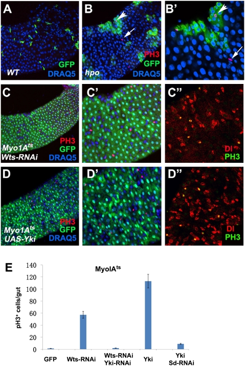
Fig. 2.
Loss of Hpo signaling can induce ISC proliferation nonautonomously. (A–B′) Adult guts containing WT (WT) control clones (A) or hpoBF33 clones (B and B′; B′ is an enlarged view of B) were immunostained to show the expression of GFP (green), PH3 (red), and DRAQ5 (blue). Control and mutant clones were generated using the MARCM system and marked by GFP expression. hpo Mutant clones stimulated cell division of neighboring WT ISCs (arrows in B and B′). Arrowheads indicate dividing cells within hpo mutant clones. (C–D′′) Adult midguts that expressed UAS-Wts-RNAi (C–C′′) or UAS-Yki (D–D′′) together with UAS-GFP using MyoIAts were immunostained to show the expression of PH3 (red in C and C′ and D and D′; green in C′′ and D′′), Dl (red in C′′ and D′′), GFP (green in C, C′, D, and D′) and DRAQ5 (blue). GFP marked ECs. Guts were dissected from adult flies grown at 29 °C for 5 d. (E) Quantification of PH3+ cells in midguts of the indicated genotypes (mean ± SD, n ≥ 15).

