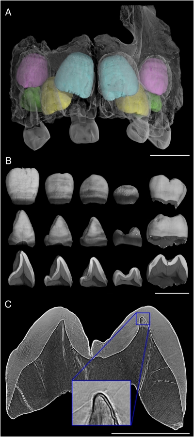
Fig. 1.
Virtual histology of the maxillary dentition from the 3-y-old Engis 2 Neanderthal. (A) Synchrotron micro-CT scan (31.3-μm voxel size) showing central incisors in light blue, lateral incisors in yellow, canines in pink, and third premolars in green. (Deciduous elements are not rendered in color as they were not studied.) (B) Isolated elements and cross-sectional slices show the degree of permanent tooth calcification and broad horizontal hypoplastic bands. (Scale bars in A and B, 10 mm.) (C) Synchrotron phase contrast image (4.95-μm voxel size) used to count long-period lines in the maxillary first molar. (Scale bar, 5 mm.) (Inset) The neonatal line just above the conical dentine horn tip of the mesiobuccal cusp, estimated to have begun forming 17 d before birth.

