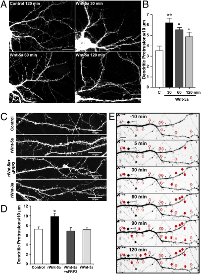
Fig. 2.
Wnt-5a increases dendritic spine density. (A) Images of dendrites of EGFP-transfected hippocampal neurons treated at DIV 12 with Wnt-5a for 30, 60, and 120 min and with control medium for 120 min. (B) Quantification of the density of dendritic protrusions versus time of treatment with Wnt-5a–conditioned medium and control medium (C). *P < 0.05; **P < 0.01. (C) Images of dendrites of DIV 14 hippocampal neurons transfected with EGFP and treated with rWnt-5a (250 ng/mL), rWnt-5a plus sFRP-2 (1 μg/mL), or rWnt-3a for 120 min. (Scale bars: 10 μm.) (D) Quantification of density of dendritic protrusions with treatments shown in C. Data represent the mean ± SE of three independent experiments; n ≥ 30 dendrite segments. *P < 0.05. ANOVA, Dunnett´s posttest. (E) Wnt-5a induces de novo formation of dendritic spines (filled arrows) and modulates preexisting spine volume (empty arrows). Live-cell time-lapse imaging of an EGFP-transfected neuron dendrite shown before (−10 min) and after 5, 30, 60, 90, and 120 min of treatment with rWnt-5a. (Scale bar: 5 μm.) (An enlarged version of the time lapse is shown in Fig. S1).

