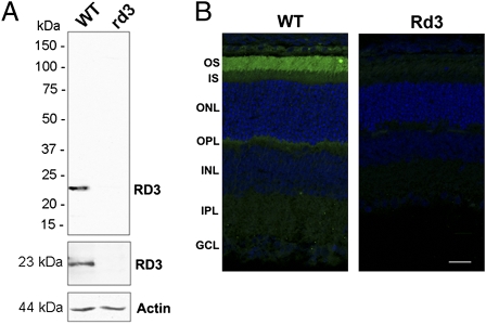
Fig. 1.
Expression and localization of RD3 in mouse retina. (A) Western blot analysis of 21-d-old WT and homozygous rd3 mouse retinal membrane extracts labeled with the Rd3-9D12 monoclonal antibody (Top), a polyclonal antibody to RD3 (Middle), and an anti-actin antibody as a loading control (Bottom). RD3 was present in the WT membrane extract, but absent in the rd3 membrane extract. (B) Confocal images of 21-d-old WT and rd3 mouse retinal cryosections labeled with the purified polyclonal antibody to RD3 (green) and counterstained with DAPI nuclear stain (blue). RD3 is localized primarily to the rod and cone outer segments. OS, outer segment; IS, inner segment; ONL, outer nuclear layer; OPL, outer plexiform layer; INL, inner nuclear layer; IPL, inner plexiform layer; GCL, ganglion cell layer. (Scale bar: 20 μm.)

