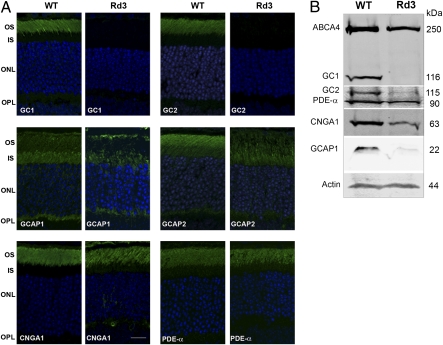
Fig. 3.
Expression and localization of GCs and other photoreceptor proteins in WT and rd3 mouse retina by immunofluorescence microscopy and Western blot analysis. (A) Retina cryosections from 21-d-old WT and rd3 mice were labeled with antibodies to GC1, GC2, GCAP1, GCAP2, CNGA1, and PDE-α. GC1 and GC2 localized to the outer segments of the WT retina but were undetectable in the rd3 retina. GCAP1 and GCAP2 localized to photoreceptor outer and inner segments of the WT retina but showed reduced expression and mislocalization in photoreceptors of the rd3 retina. CNGA1 and PDE-α localized to the outer segments of both the WT and rd3 retina. Other proteins, including ABCA4, perpherin-2, and rhodopsin, exhibited a normal outer segment distribution in the rd3 retina. (Scale bar: 20 μm.) (B) Western blots of retinal extracts from WT and rd3 mice labeled with antibodies to GC1 and GC2 and various outer segment proteins. Actin was used as a loading control.

