Fig. 5.
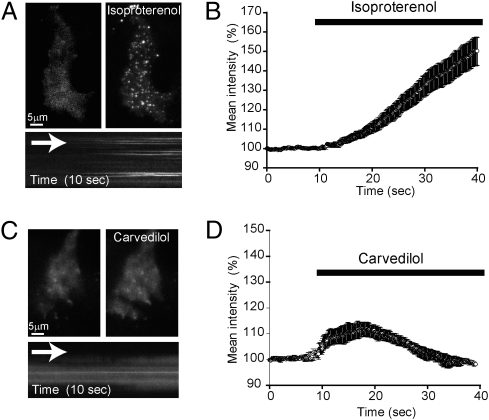
Isoproterenol and carvedilol induce distinct spatiotemporal patterns of β-arrestin recruitment to the plasma membrane. HEK 293 cells stably expressing flag-tagged β2-adrenergic receptors (∼1 pmol/mg) were transfected with β-arrestin-2–GFP and imaged by TIRFM to visualize arrestin translocation and surface distribution. (A) The addition of 10 μM isoproterenol to the incubation media induced an increase in total average fluorescence and the appearance of discrete puncta. (Lower) A kymograph from the same cell indicating the appearance of arrestin-labeled puncta. (B) Analysis of the effect of isoproterenol on mean intensity of β-arrestin-2–GFP fluorescence measured before and after bath application (n = 12 cells). (C) Application of 10 μM carvedilol to the incubation media induced an increase in surface fluorescence, indicating translocation of β-arrestin-2–GFP to the plasma membrane. Unlike isoproterenol, addition of carvedilol produced a largely diffuse surface distribution, as also shown in the kymograph. (D) Analysis of the effect of carvedilol on the mean intensity of β-arrestin-2–GFP fluorescence measured before and after bath application (n = 20 cells).

