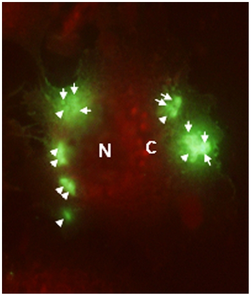
Figure 3. Multiple Pv liver stage parasites were seen in single hepatocyte in a 3 Day culture.
HepG2-A16 cells were infected with Pv spz and the liver stage trophozoites were stained with the anti-PvCSP mAb, NVS3. Some HepG2-A16 cells were seen with multiple liver stage parasites (400X magnification). N: Nucleus of hepatocyte, C: Cytoplasm of hepatocyte, White Arrows: Individual 3 Day hepatocyte stage Pv.

