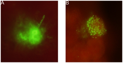
Figure 4. In-vitro development of late liver stage schizonts expressing PvMSP1.
20,000 HepG2-A16 cells were infected with 50,000 Pv spz. Three hrs later uninfected Pv spz were washed off and the culture was maintained for 9 days with daily media changes. Mature Pv liver stage schizonts (400 X magnification) in HepG2-A16 cells were stained with (A) the mAb to the PvCSP, NVS3 (20 µg/ml) or (B) with a mAb against Pv merozoite surface protein 1(PvMSP1), 3F8.A2 (1∶50 dilution). As a negative control, uninfected HepG2-A16 cells were incubated with the individual mAbs and labeled secondary antibodies. There was no evidence of staining in these negative control cultures (data not shown).

