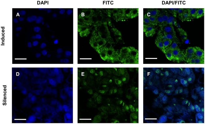
Figure 6. Presence of Cq-IAG in hAGs from endocrinologically-induced versus dsCq-IAG-injected crayfish.
Immunohistochemistry was performed on sections from the base of the fifth pereiopod of induced (top) and Cq-IAG dsRNA-injected (bottom) intersex animals. Large quantities of Cq-IAG, demonstrated by the green fluorescence of bound goat anti-rabbit FITC conjugated antibodies, were observed in the cytoplasm of induced AG cells (B, C). Reduced levels of the Cq-IAG hormone were observed in the cytoplasm of AG cells of dsCq-IAG -injected intersex animals (F). DAPI counterstain was used to identify nuclei in both induced (A, C) and silenced (D, F) intersex animals. Bar = 20 µm.

