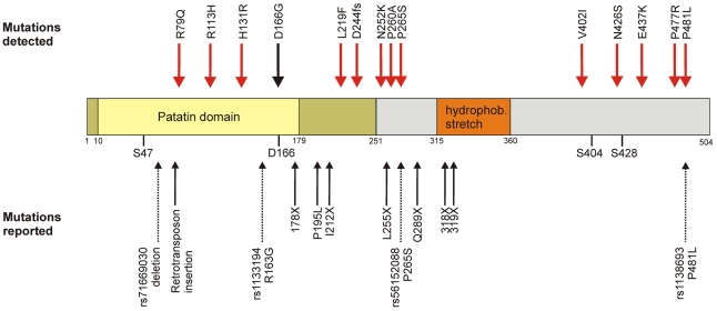
Figure 1. Domain organization and genetic variability of the ATGL protein.
Upper panel: Amino acid exchanges found in SAPHIR (red) and the patient with neutral lipid storage disease with myopathy (black). Central panel: Graphical representation of the ATGL domain organization as described in the main text. Dark yellow: α/β hydrolase fold. Yellow: Patatin domain with catalytic residues S47 and D166. Orange: hydrophobic stretch. Grey: C-Terminus. S404 and S428: phosphorylated serine residues. Lower panel: Already known variations. Bold arrows: Mutations known to cause NLSDM [41], [46]. Dotted arrows: known SNPs from dbSNP b.131.

