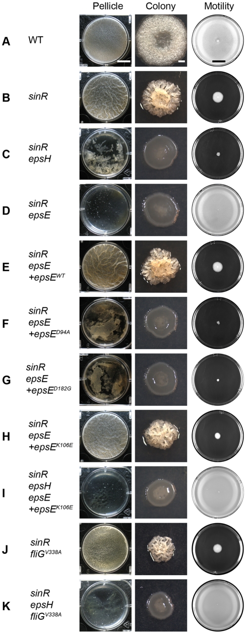
Figure 1. EpsE inhibits motility and promotes biofilm formation.
The “pellicle” column depicts top-down images of 6-well microtiter plates containing MSgg media and the indicated strains incubated for 2 days at 25°C. Scale bar equals 1 cm. The “colony” column depicts colonies grown on MSgg agar for 3 days at 25°C. Scale bar equals 1 mm. The “motility” column depicts 0.7% LB agar plates centrally inoculated and incubated overnight at 37°C. Images were taken on a black background so zones colonized agar appear white, while zones of uncolonized agar appear black. Scale bar equals 2 cm. The indicated wild type and mutant strains are as follows: A) WT 3610, B) sinR DS859, C) sinR epsH DS1674, D) sinR epsE DS2174, E) sinR epsE +epsEWT DS2239, F) sinR epsE +epsED94A DS2287, G) sinR epsE +epsED182G DS5837, H) sinR epsE +epsEK106E DS4777, I) sinR epsH epsE +epsEK106E DS5142, J) sinR fliGV338A DS3005, and K) sinR epsH fliGV338 A DS4532.

