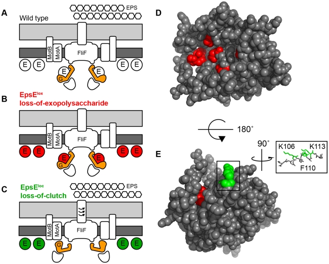
Figure 6. EpsE is a bifunctional protein that contributes to EPS biosynthesis and acts as a clutch on the flagella rotor.
A–C. Cross-section diagrams of the B. subtilis flagellar basal body. Dark grey rectangles indicate the plasma membrane while light grey rectangles indicate the peptidoglycan cell wall. “G” indicates the rotor FliG. “E” indicates EpsE. Hexagons indicate the biofilm EPS matrix. Red and green circles indicate lox and loc alleles of EpsE respectively. A) Wild type cells are capable of EPS biosynthesis and EpsE disables flagellar rotation by disrupting the FliG-MotA interaction. B) Cells containing EpsELox (Red) are incapable of EPS biosynthesis, but EpsE still acts as a clutch on FliG. C) Cells containing EpsELoc (Green) are capable of EPS biosynthesis, but are unable to interact with FliG and act as a clutch. D–E. Spacefilling model of predicted EpsE structure. Red indicates the location of lox mutations while green indicates the location of loc mutations. Inset focuses on the indicated alpha helix showing the three loc residues K106, F110, and K113 that localize to the same face.

