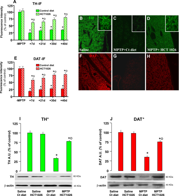
Figure 3.
HCT1026 inhibits MPTP-induced loss of striatal TH- and DAT- proteins by immunohistochemistry and western blotting. Ageing C57Bl/6 mice fed with a control (ct) or HCT1026 diets starting at -10 d, underwent an MPTP treatment, as described. At different time-intervals, mice were anesthetized and rapidly perfused, the brains were carefully removed and processed for immunohistochemistry, as described. TH- (A) and DAT-(E) IR in striatum (Str) assessed by immunofluorescent staining and image analysis by confocal Laser microscopy in ageing mice fed with ct or HCT1026 diets, treated with saline or MPTP (n = 5/time point). Fluorescence intensity values (FI, means ± S.E.M.) are expressed as % of saline. **p < 0.05 vs saline, °p < 0.05 vs MPTP fed with control diet. B-H: Representative confocal images show loss of TH-IF (revealed by FITC, green) in Str of MPTP mice fed with a ct diet at 40 dpt (C) and a substantial rescue of TH- (D) by HCT1026. F-H: Representative confocal images show loss of DAT-IF (revealed by FITC, green) in Str of MPTP mice fed with a ct diet at 40 dpt (G) and a substantial rescue of DAT-IF (H) by HCT1026. E-F: For western blot analysis, at 40 d after saline or MPTP injections in mice fed with the ct or HCT1026 diets, mice were sacrificed and striatal tissue samples processed for WB, as described. The data from experimental bands were normalized to β-actin, before statistical analysis of variance and values expressed as % of saline-injected controls, within each respective group. Note the significant decreased TH (I) and DAT (J) protein levels in MPTP mice fed with a ct diet, whereas a recovery was observed in HCT1026 fed mice. *p < 0.05 vs saline; *° p < 0.05 vs MPTP fed with ct.

