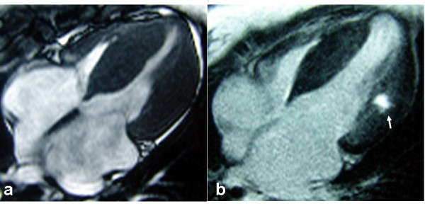
Figure 1.

MRI imaging of the case. a A four-chamber view demonstrates symmetrical thickening of interventricular septum and the lateral wall of left ventricle. b A four-chamber view shows fibrosis involving the middle layer of left ventricle lateral wall (arrow).
