Figure 2.

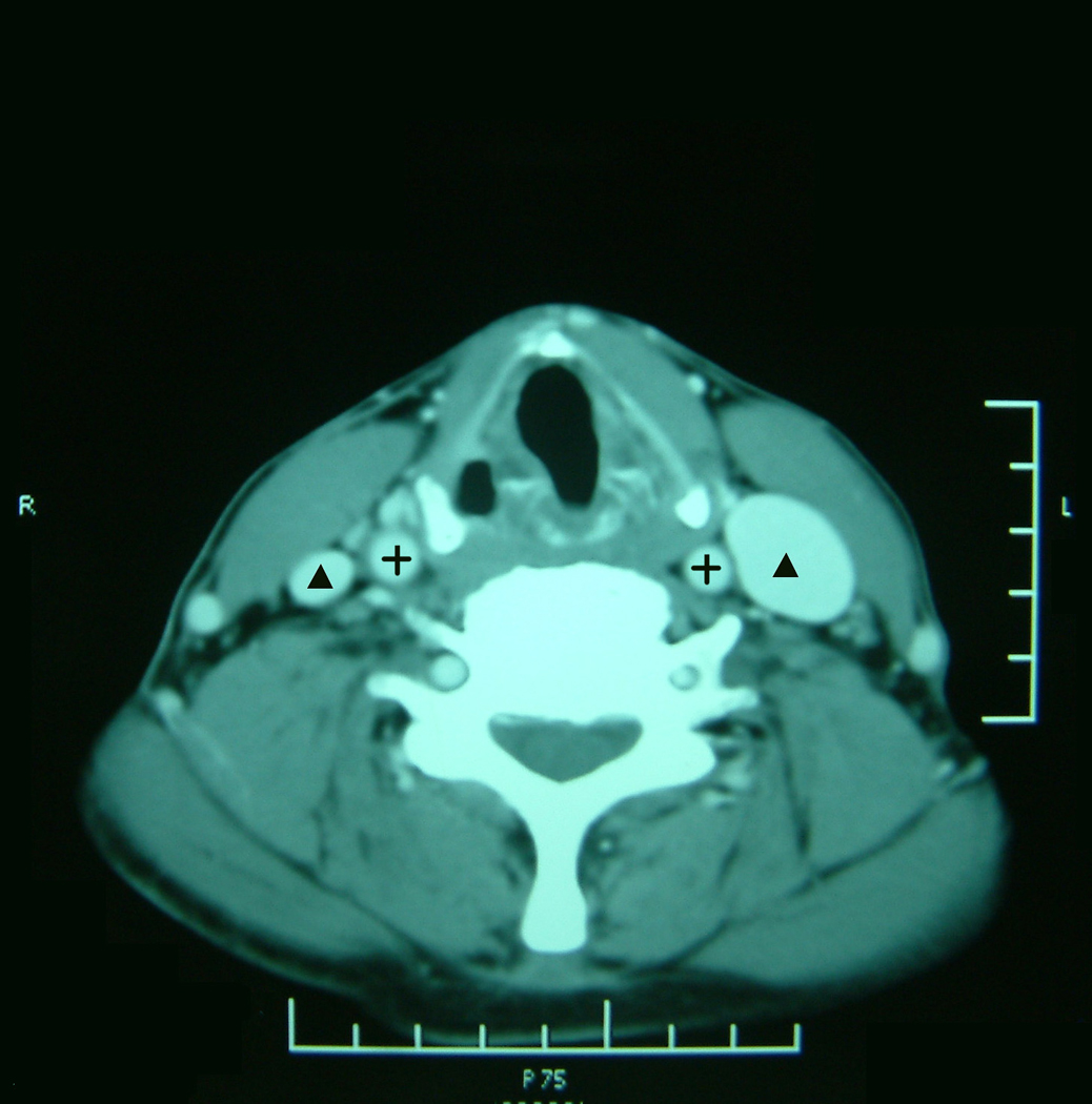
Axial contrast-enhanced CT at the level of the thyroid cartilage showing the common carotid artery (+) and the internal jugular vein (▲). The right internal jugular vein is much smaller than the left internal jugular vein.

Axial contrast-enhanced CT at the level of the thyroid cartilage showing the common carotid artery (+) and the internal jugular vein (▲). The right internal jugular vein is much smaller than the left internal jugular vein.