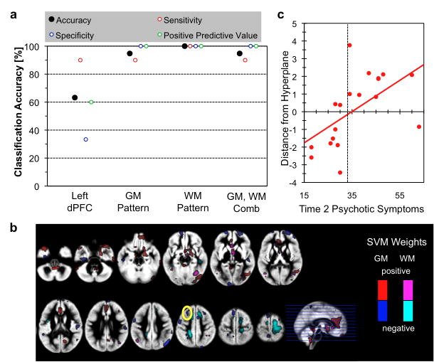
Fig 4.
Multivariate pattern analysis results. a. Classification accuracy using left PFC GM volume as single feature (left PFC), patterns of whole-brain GM volume (GM), patterns of whole-brain WM volume (WM), and combination of GM and WM (GM+WM). b. Association between distance from hyperplane for the whole-brain GM and WM pattern classifier and BPRS scores. r=0.70, p<0.001. c. Morphometric patterns that discriminate between 22q11.2DS individuals with and without psychotic symptoms. Voxels that remained during the recursive feature elimination with positive weights are plotted in red (GM) and violet (WM), and with negative weights in blue (GM) and cyan (WM). Yellow solid circle indicate overlap with univariate analysis showing associations between GMV changes and Time2 psychotic symptoms.

