
Figure 1.
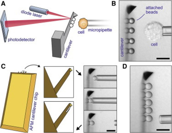
Integration of cantilever-based bead arrays into our custom-built force probe (11). (A) The conventional AFM core is turned on its side and combined with micropipetting. Cells or particles can be probed against an array of beads affixed to the cantilever. (B) Videomicrograph of a typical experiment showing a side view of the cantilever. (Reflections of the attached polystyrene beads form at the flat of the cantilever.) (C) Illustration of our procedure to assemble bead arrays on commercial AFM cantilevers. One-by-one, beads are aligned and attached to the rectangular cantilever using a micropipette. (D) Completed five-bead array; here: assembled from silica beads. (All scale bars denote 10 μm.)