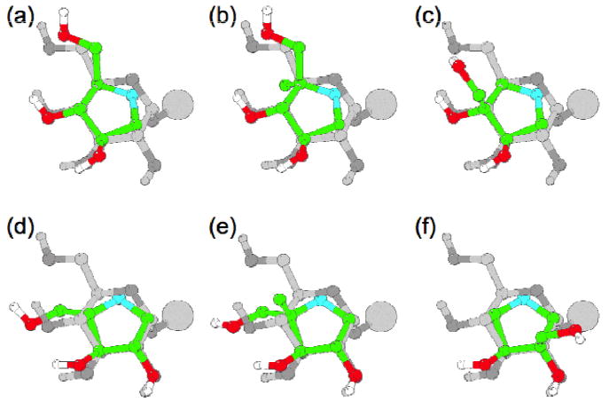
Figure 3.

Overlay of the six inhibitors, (a) DAB 3D, (b) 4-C-Me-DAB 1D, (c) isoDAB 2D, (d) LAB 3L, (e) 4-C-Me-LAB 1L and (f) isoLAB 2L, with an α-glucoside. The glucose residues are in grey, with the oxygens in slightly darker grey, and the inhibitors are in colour. Only the hydroxy protons are shown, for clarity. The large sphere shows the position of the rest of the glycan.
