
Fig.6.
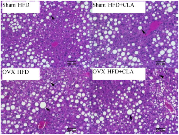
Representative pictures of H&E staining of livers after CLA treatment. Note macrosteatosis which was large fat accumulation between the central and portal vein area in Sham and OVX mice (arrow). Microsteatosis was shown in the liver of OVX mice (arrow head).