
Figure 12. CO2-dependent dye loading of the leptomeninges and astrocytes at the ventral medullary surface.
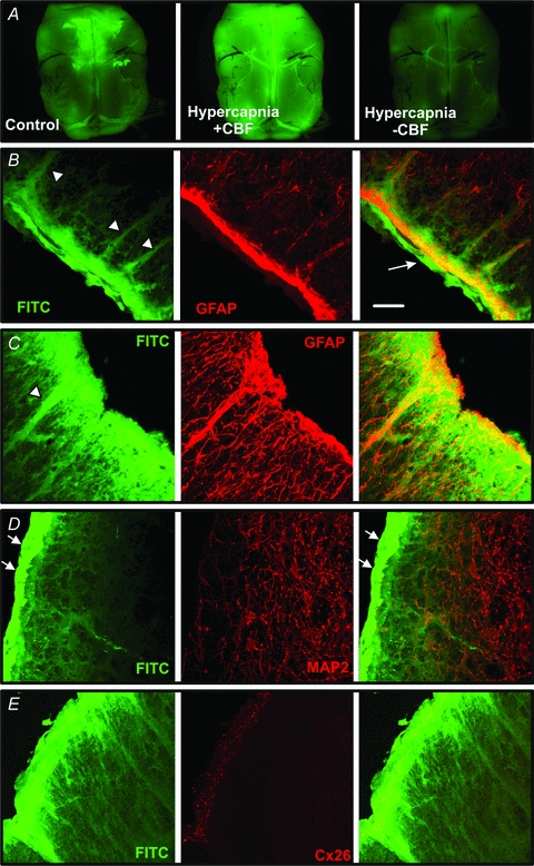
A, whole mount images of a ventral medullary slice showing baseline loading following exposure to carboxyfluorescein (CBF) in control aCSF, greatly enhanced loading when exposed to CBF during hypercapnia ( 60 mmHg) and diminished loading when
  was increased in the absence of CBF to flush out the previously loaded CBF. B–E, transverse sections showing FITC loading into the pia mater (arrow), the glia limitans and along the tracks of penetrating blood vessels (arrowheads). Note how the FITC colocalizes with GFAP staining suggesting that astrocytes are CO2 sensitive (B and C). However, neurons stained with MAP2 do not dye load with FITC. The bright strip of FITC loaded tissue (white arrows) is the leptomeningeal layer. D and E, Cx26 immunostaining is located in the glia limitans where a high degree of CO2-dependent dye loading occurs. Scale bar, 40 μm. All tissue from rat.