
Figure 1. Subpleural lymphatic vessels and experimental set-up.
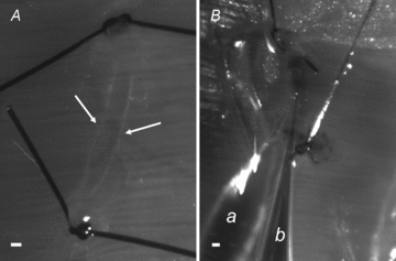
A, micrograph taken under white light epi-illumination of a lymphatic segment delimited by two occluding silk knots positioned at a distance of about 3300 μm along a linear submesothelial lymphatic vessel. Lymphatic vessels appear as darker-than-background conduits delimited by faint white borders (arrows). Underneath the lymphatic vessel, the diaphragmatic muscular fibres can be distinguished crossing the vessel almost perpendicularly. B, micrograph showing the simultaneous insertion of two micropipette tips into the lumen of a lymphatic segment prepared in a vessel belonging to a complex loop. Only the tract used for the measurements was ligated. a, micropipette for Plymph recording; b, micropipette for bolus injection. Scale bars: 200 μm.