
Figure 7. Three-dimensional modelling of the lymphatic vessel wall.
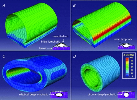
Stress distribution maps obtained through finite element modelling of ideal diaphragmatic initial lymphatics located (as depicted in the schematic drawing at the bottom right of each panel): A, immediately beneath the mesothelium. The ellipsoidal superficial vessel is delimited mostly by a thin wall of lymphatic endothelium plus pleural mesothelium and lays on a diaphragmatic muscular/tendineous support; B, deeper in the submesothelial tissue. This intermediate vessel is only partially delimited by a thin wall, most of the lateral surface being surrounded by the muscular/tendineous tissue; C, among the diaphragmatic muscular/tendineous fibres surrounded by an isotopic tissue and with a ellipsoidal cross-sectional area; D, among the diaphragmatic muscular/tendineous fibres surrounded by an isotopic tissue and with a circular cross-sectional area. Modelling was performed using the geometrical parameters presented in Table 1 and by loading the vessels with an intraluminar distending pressure of 10 mmHg. The circumferential stress distribution was identified by colours (see key) on a scale from blue (low stress) to red (high stress); the thin lines delimit the shell and solid elements used to model the thin mesothelial/endothelial wall and the solid muscular/tendineous tissue, respectively. Superficial vessel tensile stress was higher in submesothelial superficial (A) and intermediate (B) vessels which underwent the greatest deformation. In deeper vessels (C and D) surrounded by stiffer tissue, wall tension was lower and more homogenously distributed over the entire surface, particularly in perfectly circular model (D). According to these models, the mechanical properties and the function of initial lymphatics may depend upon surrounding tissue stiffness: submesothelial lacunae delimited by a complaint wall serve as reservoirs of drained fluid, while deeper vessels running the stiff tissue more efficiently propel fluid along the network.