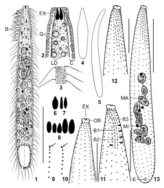
Figs 1 – 13.
Suturothrix monoarmata, Singapore (1, 3, 6, 12, 13), Thailand (2, 4, 5, 7, 9 – 11), and Brazil (8) specimens from life (1 – 8) and after protargol impregnation (9 – 13). 1: Right side overview. 2: Semischematic view of oral region. 3: Cortical granulation. 4, 5: Contracted (80 μm) and extended (120 μm) specimen. 6 – 8: Oral bulge extrusomes. 9: Anterior region of ciliary rows with supposed dikinetids marked by arrows. 10, 11: Ventral and dorsal view of same specimen. Arrowheads mark monokinetids between dikinetids. 12, 13: Dorsal ciliary pattern and nuclear apparatus of holotype specimen. B, B1–3: dorsal brush (rows), C: cortex, E: excretory pore; EX: extrusomes; G: cortical granules; LD: lipid droplets; MA: macronucleus; MI: micronucleus; OB: oral bulge. Bars 10 μm (Figs 10 – 12), 20 μm (13), 30 μm (1).

