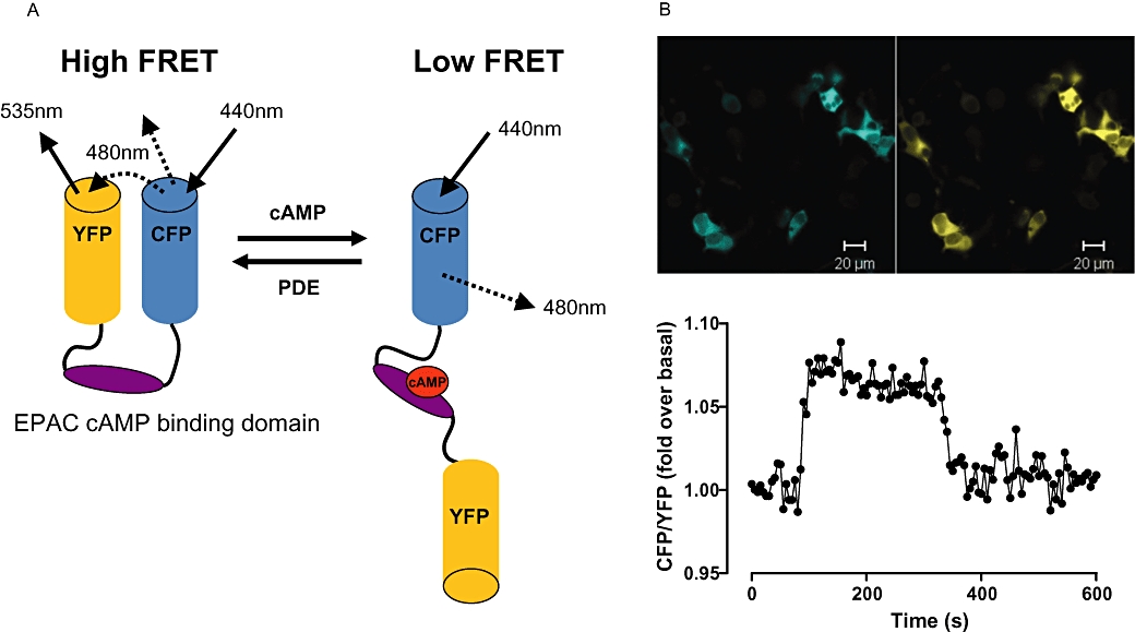
Figure 2.

EPAC-based cAMP FRET sensor. (A) A schematic showing the basic design of an EPAC-based cAMP FRET sensor. In the absence of cAMP the conformation of the sensor is such that the fluorophores are in close proximity, generating FRET. Upon cAMP binding, conformational changes result in a decrease in FRET. (B) Top panel: Fluorescent images of HEK293T cells transiently transfected with a CFP/YFP FRET sensor containing the EPAC2 cAMP binding domain. CFP and YFP fluorescence are shown in blue and yellow respectively. Lower panel: A single cell trace plotted as a ratio of CFP: YFP intensity from HEK293T cells transiently tranfected with the EPAC2-based cAMP sensor. Exposure of cells to 5′-N-ethyl carboxamide adenosine at 30 s stimulates cAMP accumulation through an endogenously expressed adenosine A2B receptor. Inhibition of cAMP accumulation results from the addition of the adenosine receptor antagonist xanthine amine congener at 300 s. CFP, cyan fluorescent protein; EPAC, exchange protein directly activated by cAMP; FRET, fluorescence resonance energy transfer; PDE, phosphodiesterase; YFP, yellow fluorescent protein.
