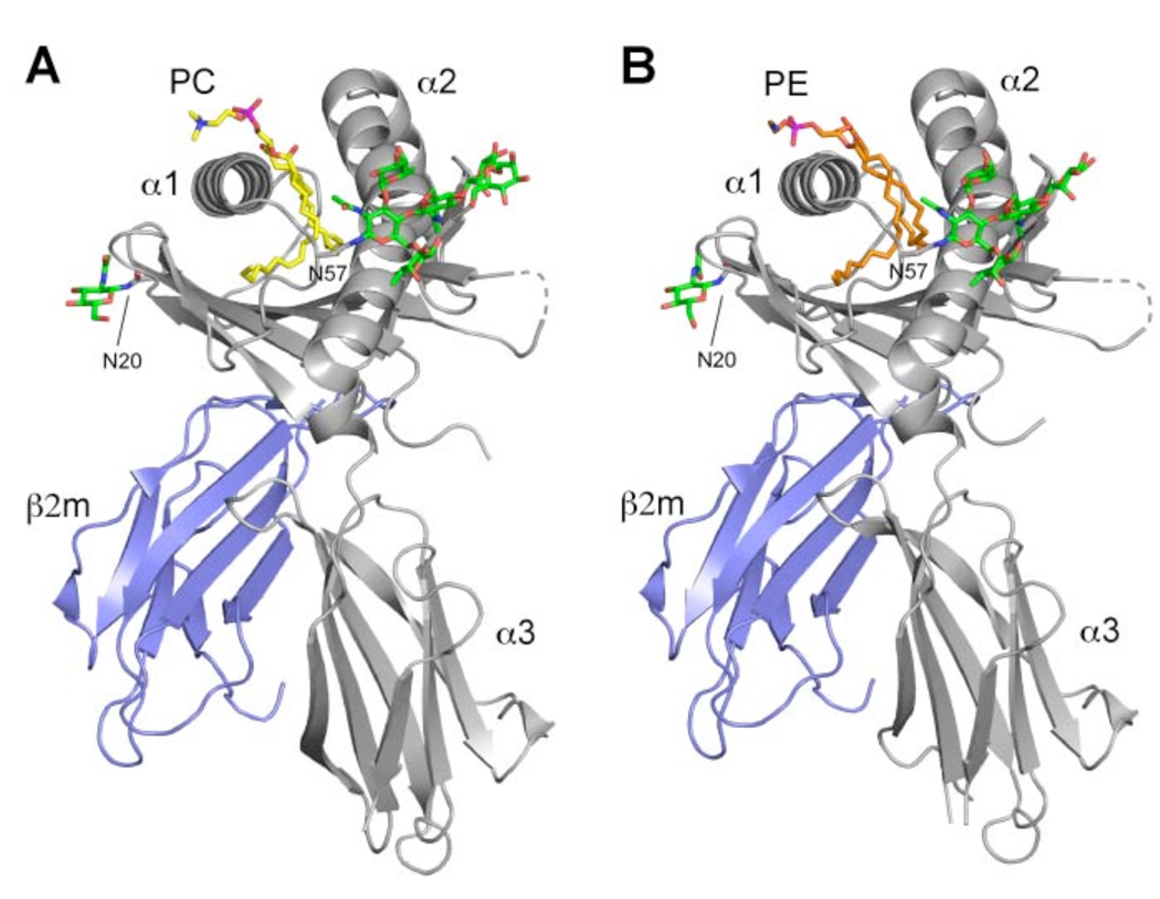
Figure 1.
Overview of the boCD1b3-PC/PE structure. CD1 heavy chain is in grey with the α1, α2 and α3 domains indicated and β2m is in blue. The bound PC molecule (A) is shown in yellow and PE in orange (B), while N-linked oligosaccharides at positions Asn20 and Asn57 are shown in green. The disordered loop comprising residues 106–109 of the heavy chain is shown as a dashed line.

