Figure 4.
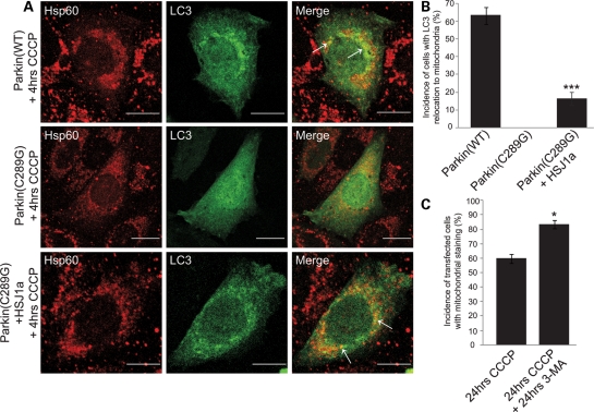
HSJ1a rescues Parkin(C289G) for mitophagy. (A) SK-N-SH cells co-transfected with FLAG-Parkin(WT) or FLAG-Parkin(C289G), myc-HSJ1a(WT) and GFP-LC3 (green) were treated with CCCP (20 µm, 4 h) and immunolabeled with anti-Hsp60 (red). (B) The percentage of cells with overlapping GFP-LC3 and MitoDsRed staining was quantified in cells co-transfected with FLAG-Parkin(WT), FLAG-Parkin(C289G) ± myc-HSJ1a(WT) and treated with CCCP (20 µm, 4 h). Error bars: ±2 SE, n = 4, ***P < 0.001. (C) Quantification of the incidence of transfected cells with mitochondrial staining in cells treated with CCCP (20 µm, 24 h, left bar), CCCP + 3-MA (10 mm, 24 h, right bar). Error bars: ±2 SE, n = 4, *P < 0.5.

