Figure 8.
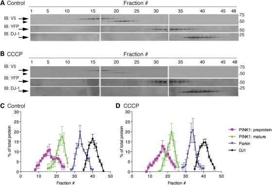
Lack of formation of co-complexes of DJ-1 and PINK1/parkin. (A and B) Size exclusion chromatography was performed on cell lysates from M17 lines stably transduced with V5-tagged PINK1 and transfected with myc-parkin either without (A) or after treatment with CCCP (B). Fractions (0.25 ml) were taken and run on SDS–PAGE gels then blotted for V5 for PINK1 (upper blots), myc-parkin (middle blots) or DJ-1 (lower blots). Markers on the right indicate sizes on SDS–PAGE in kilodaltons. (C and D) Quantification of proteins in distinct native complexes for blots as in (A) and (B) for the preprotein of PINK1 (purple), the mature PINK1 protein (green), parkin (blue) or DJ-1 (black). For each protein, the immunoreactivity in each fraction is plotted as a percentage of the total protein immunoreactivity in all fractions against fraction number. Error bars indicate SEM from n = 3 experiments from independent transfections on different occasions.

