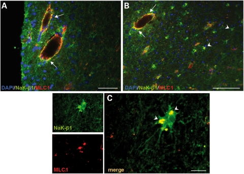
Figure 6.
Co-immunolocalization of NaK-β1 and MLC1 in multiple sclerosis (MS) brain lesions. (A and B) Immunofluorescence staining of MS brain tissue with anti-MLC1 pAb (red) and anti-Na,K-ATPase β1 (NaK-β1) mAb (green) shows that MLC1 and NaK-β1 immunoreactivities partially overlap around inflamed blood vessels (A and B, arrows) and in hypertrophic astrocytes in the lesioned periventricular white matter (B, arrowheads). (C) High-power magnification of a confocal image shows the overlapping vesicular localization of MLC1 and NaK-β1 in reactive astrocytes (arrowheads). Scale bars: A: 50 µm; B: 100 µm; C: 10 µm.

