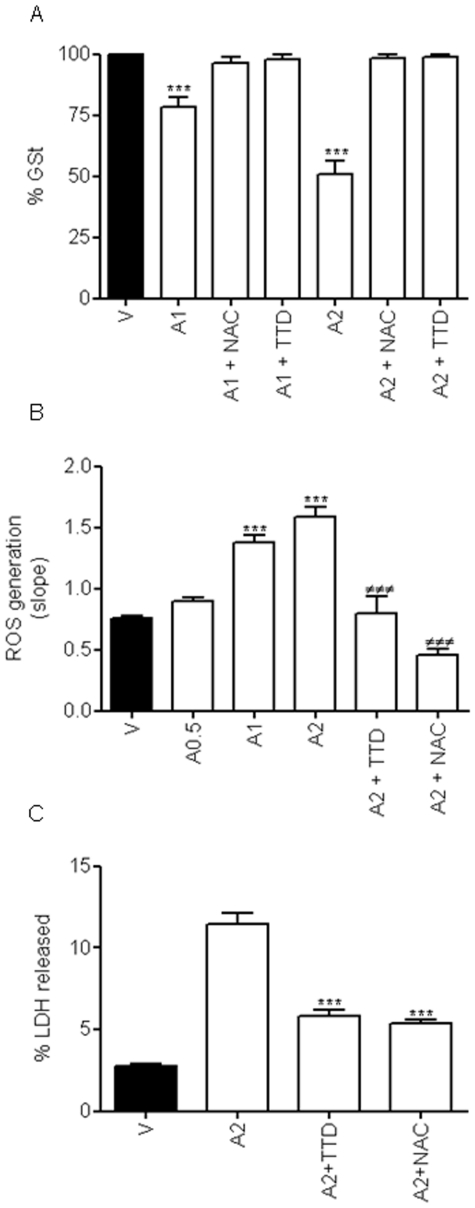
Figure 5. Redox status of rat cortical neurons.
(A) Neuronal glutathione levels were decreased by treatment with 1 mM (A1) or 2 mM (A2) AAP in a concentration-dependent manner whereas co-treatment with either N-acetylcysteine (NAC; 100 µM) or disulfiram (TTD; 0.1 µM) completely restored glutathione content to basal levels. V stands for vehicle (DMSO 1‰)-treated cells. Data represent mean ± SEM of 9 independent experiments. *** p<0.001 as compared to vehicle-treated cells). (B) AAP 0.5 (A0.5), 1 (A1) and 2 mM (A2) induced a dose-dependent increase in the rate of reactive oxygen species (ROS) production, that was prevented by both N-acetylcysteine (NAC; 100 µM) and disulfiram (TTD, 0.1 µM). V stands for vehicle (DMSO 1‰)-treated cells. Data represent the mean ± SEM of 9 independent experiments. ***, p<0.001 as compared to vehicle-treated cells (V); ###, p<0.001 compared to AAP 2 mM (A2). (C) NAC (100 µM) as well as TTD (0.1 µM), by reducing free radical production, markedly reduce AAP-mediated toxicity on rat cortical neurons. Cortical neurons were incubated with vehicle (DMSO 1‰; V) or AAP 2 mM (A2) alone or in the presence of NAC 100 µM (A2+NAC) or TTD 0.1 µM (A2+TTD) for 24 h and percentage of LDH released to the culture medium was determined. Data represent the mean ± SEM of 9 independent experiments. ***, p<0.001 as compared to AAP 2 mM-treated cells.

