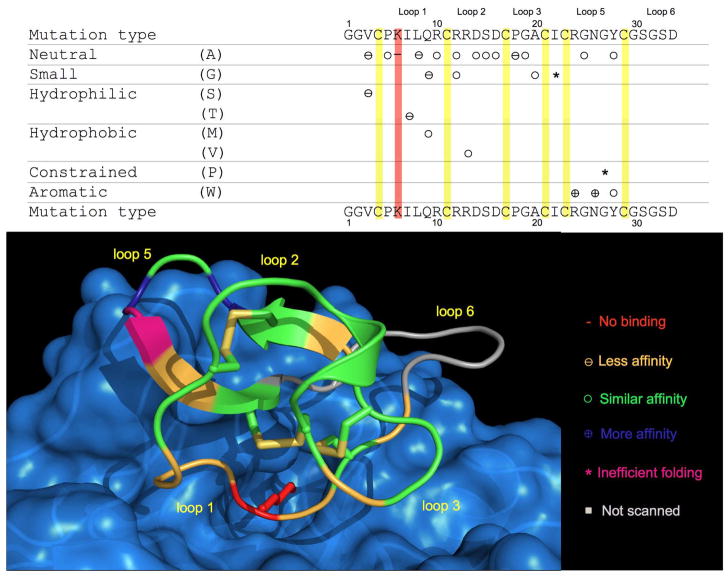
Figure 3.
Relative affinities for trypsin of a series of MCoTI-I mutants covering all the loop positions except loop 6 and Cys residues. A model of cyclotide MCoTI-I bound to trypsin is shown at the bottom indicating the position of the mutations. The side-chain of residue K6 is shown in red bound to specificity pocket of trypsin. The model was produced by homology modeling at the Swiss model workspace41 using the structure of CPTI-II-trypsin complex (PDB code: 2btc)42 as template. Structure was generated using the PyMol software package. Figure adapted from reference.16

