Fig. 4.
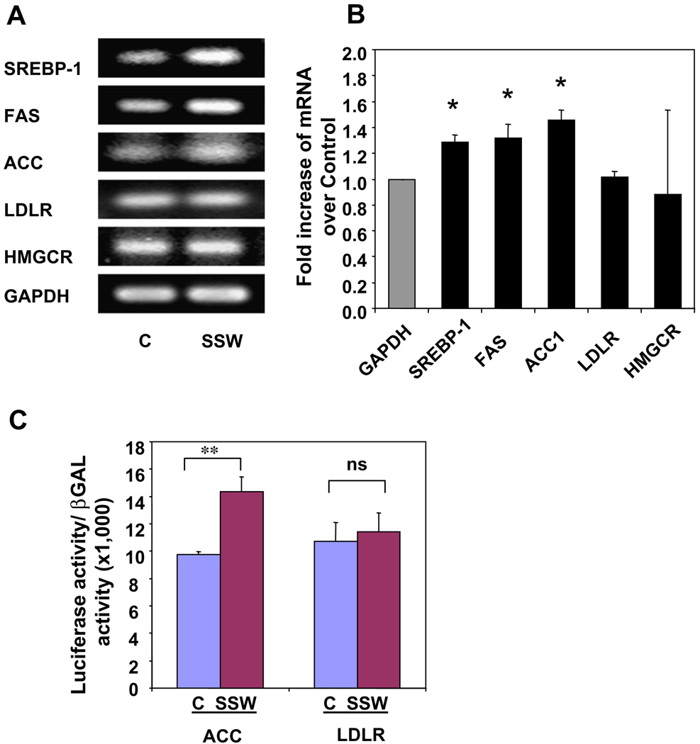
SSW-induced SREBP-1 activation, and expression of down stream genes. (A–B) Gene expression of molecules down-stream of SREBPs. The AML 12 cells were treated with 1:40 dilution of SSW or fresh media for 8 h, and total RNA extracted. Gene expression for SREBP-1, FAS, ACC, LDLR, HMGCR, and GAPDH was analyzed by RT-PCR (A) and by quantitative real-time PCR (B). (C) AML12 cells cultured in 12-well plates were transiently transfected with LDLR-Luc (1.0 µg/well) or ACC-Luc (1.0 µg) plus β-gal (0.1 µg). Thirty-six hours after transfection, the cells were treated with 1:40 dilution SSW or fresh media for 12 h. Luciferase activity was measured and normalized to that of β-gal. All of the values presented here represent a ratio of luciferase to β-Gal to correct for nonspecific variations in the transfection and assay procedures. The data represent the ratio of luciferase activity (relative light units) for the indicated test plasmid to the β-galactosidase activity (OD 420 nM/h) expressed from the control cytomegalovirus promoter. The average of at least five independent experiments for each plasmid performed in duplicate is presented.

