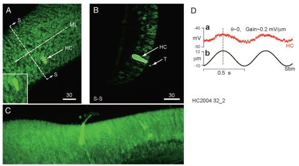
FIG. 4.
Characterized and labeled hair cell. Cells were characterized physiologically using mechanical stimuli, injected, and visualized using streptavidin-Alexafluor 568 and multiphoton microscopy. A: maximum intensity projected image showing the location of an injected hair cell (HC) in the central region of the crista (scale bars: 30 μm). Approximate locations of the midline (ML) and center (SS) are indicated. Background staining is biotin, normally present in the cells, and recognized by the streptavidin. B: a single optical slice showing the same hair cell viewed in the direction SS perpendicular to the ML. C: a rotated, rendered maximum intensity projected image in which the presence of tracer in the hair bundle is clear. D: response of the same cell to a ±10-μm sinusoidal mechanical indentation (b) of the canal duct at 2 Hz. Voltage modulation of this cell in current clamp (a) was in phase with the stimulus and the gain was 0.2 mV/μm indent.

