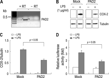
FIGURE 2.
Suppression of NF-κB activity by PAD2 overexpressed in RAW 264.7 cells. A, RT-PCR for PAD2. Expression of PAD2 mRNA was determined by PCR with two sets of primers amplifying 277 bp (*; lane 2) and 246 bp (**; lane 3) PAD2 cDNA fragments. Total RNA extracted from untreated cells was used. PCR without RT reaction (−RT) served as negative controls. PCR products were sequenced to confirm the specificity of the products. B, effect of PAD2 overexpression on COX-2 production. Cells transfected with the full-length mouse PAD2 were treated with LPS (1 μg/ml) for 4 h. COX-2 production was determined by immunoblot. Cells transfected with vector only (Mock) served as controls. Tubulin served as an internal control for immunoblot. C, comparison of COX-2 expression between control cells and PAD2-expressing cells. Densitometric measurements of immunoreactive bands were done (n = 3). The intensity of COX-2 was normalized to the intensity of tubulin. D, NF-κB activity in control cells and PAD2-expressing cells. Cells transfected with the NF-κB reporter plasmid and PAD2 or none were incubated for 24 h and then treated with LPS for 4 h. NF-κB activity was measured by luciferase assay (n = 4) as described previously (22). The activity was presented relative to the value in mock cells without PAD2 and LPS (leftmost bar).

