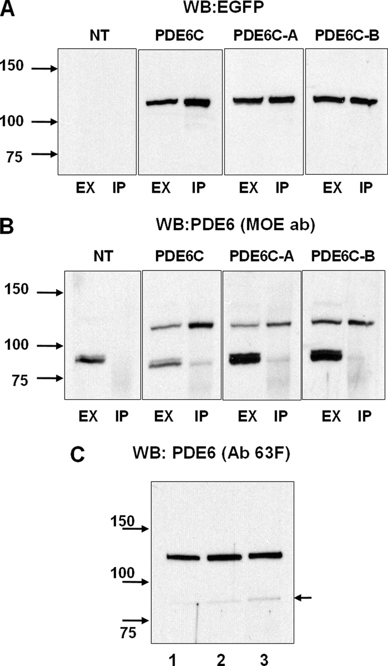
FIGURE 2.

Immunoprecipitation of EGFP-PDE6C, EGFP-PDE6C-A, and EGFP-PDE6C-B. Extracts (EX) and immunoprecipitates with sheep anti-GFP antibodies (IP) from retinas of non-transgenic (NT) and transgenic X. laevis were immunoblotted with anti-GFP monoclonal antibody B-2 (A), stripped, and reprobed with anti-PDE6 antibody (ab) MOE (B). C, immunoprecipitates of PDE6C (lane 1), PDE6C-A (lane 2), and PDE6C-B (lane 3) with sheep anti-GFP antibodies were immunoblotted with anti-PDE6 antibody 63F. The level of coprecipitation (co-dimerization) with frog PDE6AB (indicated by the arrow) was <3%. WB, Western blot.
