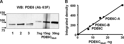
FIGURE 3.
Solubilization and quantification of PDE6C, PDE6C-A, and PDE6C-B. A, the beads with bound EGFP-PDE6C (lane 1), EGFP-PDE6C-A (lane 2), and EGFP-PDE6C-B (lane 3) were treated with trypsin, and proteins released into the soluble fraction were analyzed by immunoblotting with anti-PDE6 antibody (Ab) 63F. Purified recombinant PDE6C-His6 was used as the standard. WB, Western blot. B, PDE6C, PDE6C-A, and PDE6C-B were quantified by measuring the integrated densities of the corresponding bands and PDE6C-His6 bands corrected for the background using ImageJ.

