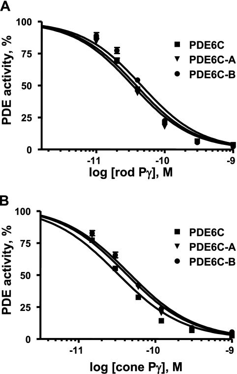
FIGURE 5.
Inhibition of PDE6C, PDE6C-A, and PDE6C-B by the cone and rod Pγ subunits. Various concentrations of rod Pγ (A) and cone Pγ (B) were added to trypsin-released PDE6C, PDE6C-A, and PDE6C-B (1 pm each). The Ki values for inhibition with rod Pγ are as follows: PDE6C, 35 ± 5 pm; PDE6C-A, 38 ± 4 pm; and PDE6C-B, 46 ± 6 pm. The Ki values for inhibition with cone Pγ are as follows: PDE6C, 33 ± 4 pm; PDE6C-A, 40 ± 5 pm; and PDE6C-B, 45 ± 4 pm. Results from one of three similar experiments are shown.

