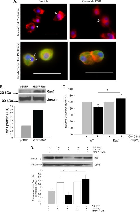
FIGURE 6.
Ceramide inhibits AM efferocytosis through Rac1 down-regulation. A, representative fluorescence micrographs of AM (either alone in the upper panels or co-incubated with apoptotic Jurkat cells in the lower panels) stained for actin (with Texas Red phalloidin; red), nuclear marker (DAPI; blue), and Rac1 (with Rac1 antibody conjugated to Alexa fluor 488; green; lower panels only) following treatment with Cer C6:0 (10 μm; 4 h) or control vehicle (0.1% ethanol). Note that control cells exhibit ruffles of the plasma membrane (arrows), whereas ceramide-treated cells have a near loss of membrane ruffle formation (double arrowhead) and decreased Rac1 staining. The engulfed Jurkat apoptotic cells are seen in control cells (asterisk), surrounded by a Rac1-rich phagosome membrane (arrow). Scale bar, 50 μm. The values are representative of n = 2 experiments. B, Rac1 protein level in NR8383 cells transfected with constitutive active Rac1 or control plasmid. Vinculin immunoblot was used as loading control. C, phagocytic index of wild type NR8383 macrophages and those overexpressing Rac1 treated with ceramide (Cer C6:0; 10 μm; 4 h); note the lack of inhibitory effect of ceramide on efferocytosis in cells expressing a constitutively active Rac1 (means ± S.E.; n = 3). D, Rac1 plasma membrane abundance detected in protein lysates from total membrane fractions obtained from NR8383 cells treated with CS extract (3%; 4 h) or AC with or without a ceramidase inhibitor pretreatment (MAPP; 1 μm, 2 h). The proteins were detected by Western blotting using a specific Rac1 antibody, CD71 was used as loading control; densitometry of Rac1 expression was normalized by loading control (means ± S.E.; n = 3; *, p < 0.05).

