
Figure 1.
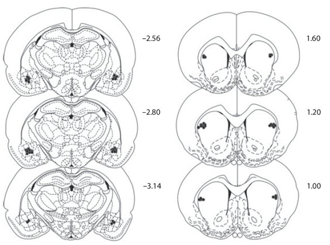
Schematic diagram of coronal sections (anterior–posterior from bregma) of infusion cannulae placements for the basolateral amygdala (left) and the dorsolateral caudate putamen (right) (adapted from Paxinos & Watson, 1997).
Official websites use .gov
A
.gov website belongs to an official
government organization in the United States.
Secure .gov websites use HTTPS
A lock (
) or https:// means you've safely
connected to the .gov website. Share sensitive
information only on official, secure websites.
Schematic diagram of coronal sections (anterior–posterior from bregma) of infusion cannulae placements for the basolateral amygdala (left) and the dorsolateral caudate putamen (right) (adapted from Paxinos & Watson, 1997).