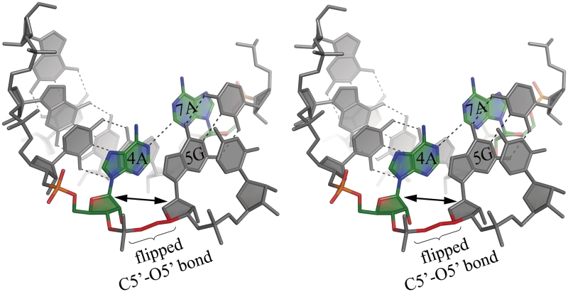
Figure 1.
Stereo view of the A–A pair (green) and its surroundings in the (GGCAGCAGAA)2 0.95 Å resolution structure. The α- and γ-backbone torsion angles between A4 and G5 take unusual values (see text) which results in local unwinding of the helix. This can be seen by comparing the orientation of consecutive ribose rings, which appear co-planar in the two residues. This conformation of the backbone is found in all the examined A–A pairs and is associated with the adenosine less inclined towards the major groove.

