Figure 4.
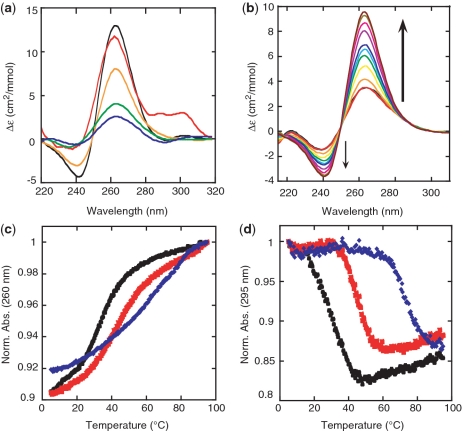
Formation of GQS RNA oligonucleotides (a) CD spectra of RNA GQS in 10 mM LiCacodylate (pH 7.0) and 150 mM KCl at 20°C: G3L221 (black), G2L111 (red), G3L444 (gold), G3L444 + FLANK (green) and G2L444 (blue). The positive peak at 260 nm and the negative peak at 240 nm suggest that the RNA GQS adopt a parallel conformation. The G2L111 RNA most likely has some antiparallel character due to the shoulder in the spectrum extending to 300 nm. See ‘Materials and Methods’ section for full sequences. (b) Sample K+ titration. Titration is of G3L444 RNA GQS with KCl additions from 0 mM to 700 mM. The arrows indicate increasing K+ concentrations. Also included are UV thermal denaturations of G3L444 RNA in 100 mM LiCl (black), NaCl (red) and KCl (blue) at (c) 260 nm and (d) 295 nm. Absorbances are normalized to the highest absorbance.

