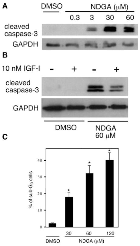
Fig. 5.
NDGA causes caspase activation and apoptosis in neuroblastoma cells. A: SH-SY5Y cells grown in serum containing-medium were treated with DMSO or 0.3–60 μM NDGA for 12 h. Activated caspase-3 fragments were detected using Western blot analysis. Upper panel shows the 14/17 kDa cleavage fragments of caspase-3, while the lower panel shows GAPDH expression as a loading control. A representative of three separate experiments is shown. B: Serum-starved SH-SY5Y cells were treated with DMSO or 60 μM NDGA for 12 h. Some cultures included 10 nM IGF-I for the entire treatment period. Lysates were collected and caspase-3 cleavage fragments were detected as above. C: SH-SY5Y cells were treated with DMSO or NDGA (30–120 μM) for 24 h, fixed, stained with propidium idodide, and subjected to flow cytometric analysis of cell cycle. Bars represent the mean ± SEM percentage of cells in the sub-G0 apoptotic phase from five separate experiments. *P <0.05 versus DMSO.

