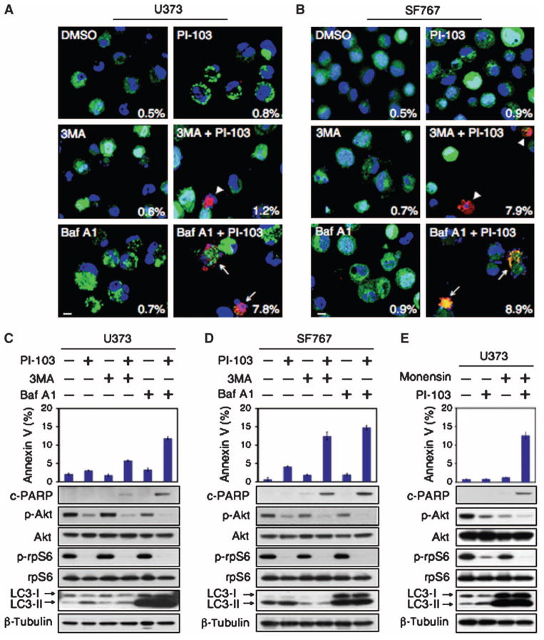
Fig. 2.
PI-103 synergizes with Baf A1, an inhibitor of autophagosome maturation, to induce apoptosis in glioma. Glioma cells (U373, PTEN mt and SF767, PTEN wt) were transduced with pBabe-GFP-LC3, treated with DMSO or PI-103 (1 μM) for 24 hours, and then treated for 48 hours with either 3MA (5 mM) or Baf A1 (10 nM). (A and B) Cytospin preparations were stained with antibody against cleaved caspase 3 (red) or GFP-LC3 (green). Nuclei stained in blue (Hoechst dye). Arrowheads indicate cleaved caspase 3–positive cells, with percentages indicated. Arrows show cleaved caspase 3–positive cells with condensed GFP-LC3 punctate dots. Scale bar, 100 μm. (C and D) Apoptotic cells were analyzed by flow cytometry. Percentages of cells positive for annexin V are the mean ± SE for triplicate samples (U373: P < 0.0001 by Student’s t test for PI-103 plus 3MA versus DMSO; P < 0.0001 for PI-103 plus Baf A1 versus DMSO; SF767: P < 0.0001 by Student’s t test for PI-103 plus 3MA versus DMSO; P < 0.0001 for PI-103 plus Baf A1 versus DMSO). Lysed cells were analyzed by immunoblot with antisera indicated. (E) To exclude off-target effects of Baf A1 independent of lysosomal trafficking, we treated U373 PTEN mt glioma cells with 1 μM PI-103 or DMSO for 24 hours. Monensin (3 μM) was added where indicated (24 hours), and cells were analyzed by flow cytometry for annexin V. Data show error among triplicate measurements for each value (top panel). An aliquot of cells was analyzed by immunoblot with antisera indicated (bottom panel).

