
Fig. 1.
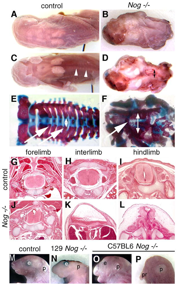
Spina bifida and exencephaly in Nog−/− pups. (A–D) Whole-mount dorsal views of perinatal pups. (A) Wild-type pup Nog+/− intercross, 129/Sv genetic background. (B) Nog homozygous littermate showing exencephaly and spina bifida, covered by skin. (C) Wild-type pup with dorsal skin removed. Arrowheads point out neural arches around spinal cord. (D) Nog homozygote with skin removed, showing open neural tube and gap between neural arches in lumbar region (doubleheaded arrow). (E and F) Skeletal preparations stain bone in red and cartilage in blue. Wild-type pup (E) shows a regular pattern of neural arches (arrows) converging over the neural tube (doubleheaded arrow). (F) Nog−/− pup with dysmorphic neural arches (arrow), and wide distance between spinal sides (doubleheaded arrow). Note general dysplasia of axial skeleton. (G–L) Histological transverse sections of wild-type and Nog late gestation littermates. (G–H) In control fetuses, a closed neural tube and orderly arrangement of skeletal and muscular tissues are seen at all axial levels. (J) Relatively normal neural tube histology in Nog−/− pups at the level of the forelimb. (K) Severe defects in Nog−/− between the limbs, where the neural tissue appears as a thickened ventral plate under a broad lumen overlain by skin and connective tissue: spina bifida occulta. (L) The neural tube at the level of the hindlimb shows less severe defects in the mutant. (M–P) Craniofacial defects in Nog mutants vary with genetic background. Lateral whole-mount views. (M) Wild-type pup. (N) Nog−/− in 129/Sv background with exencephaly. (O) Nog−/− pup in C57BL/6 background with mild facial truncations. (P) Rare Nog−/− pup in C57BL/6 with severe craniofacial truncation. p, pinna (external ear); pr, proboscis.